
CAT #: IGK Clonality
IGK Clonality Assay
CONTACT US ⟶-
Description of Test:
For detection of the vast majority of IGK gene rearrangements, a multiplex master mix targeting the conserved Jƙ-Cƙ intron, Kde, and Jƙ regions is used for PCR amplification. Next-generation sequencing of the PCR products is used to identify DNA sequences specific to clonal gene rearrangements. Bioinformatics tools facilitate the characterization of sequences present at greater than 5% of the population. These sequences can be used to track specific clonal populations.
-
Overview:
During development of lymphoid cells, antigen receptor genes undergo somatic gene rearrangements.1
The human immunoglobulin kappa (IGK) locus on chromosome 2 (2p11.2) includes 7 Vƙ (variable region) gene segments and 5 Jƙ (joining region) gene segments upstream of the Cƙ region. The kappa deleting element (Kde), approximately 24 kb downstream of the Jƙ-Cƙ region, can also rearrange with Vƙ gene segments and the isolated recombination signal sequence in the Jƙ-Cƙ intronic region.2
Specifically, during B-cell development, genes encoding IGK molecules are assembled from multiple polymorphic gene segments that undergo rearrangements generating gene receptors unique in both length and sequence. Since leukemias and lymphomas originate from the malignant transformation of individual lymphoid cells, all leukemias and lymphomas generally share one or more cell-specific or “clonal” antigen receptor gene rearrangements. Therefore, tests that detect IGK clonal rearrangements can be useful in the study of B- and T-cell malignancies.
CLINICAL UTILITY
Immunoglobulin kappa light chain (IGK) clonality studies are often used in conjunction with immunoglobulin heavy chain (IGH) clonality studies to distinguish B-cell neoplasm from reactive B-cell proliferation or reactive plasmacytosis. They are particularly useful under the following conditions:
• A suspected/equivocal clonal B-cell population is detected by flow cytometry, and clonality needs to be confirmed;
• Atypical lymphoid infiltrate is present on the tissue sections (lymph nodes or extra nodal tissue) based on morphology review, and immunophenotypic studies by flow cytometry are not available and/or no obvious immunophenotypic aberrancy can be demonstrated by immunohistochemical stains;
• Dense plasmocytic infiltrate on the tissue sections (lymph nodes, bone marrow clot or other extra nodal tissue) based on morphology review, and
− immunophenotypic studies by flow cytometry are not available to evaluate the plasma cells and/or B-cells,
− no obvious clonal plasma cell population can be identified by immunohistochemical stains or in situ hybridization, and
− no obvious immunophenotypic aberrancy can be demonstrated on the plasma cells and/or B-cells by immunohistochemical stains;
• Investigating the clonal relationship between the current specimen with a diagnosis of B-cell lymphoma/leukemia and a prior specimen with a diagnosis of B-cell lymphoma/leukemia.
While IGH and/or IGK clonality studies are essential for the differential diagnosis of B-cell lymphoma vs. reactive B-cell proliferations sometimes, it is important to understand that the diagnosis cannot be based on molecular studies alone but requires careful correlation of clinical, morphologic, immunophenotypic and genetic features.
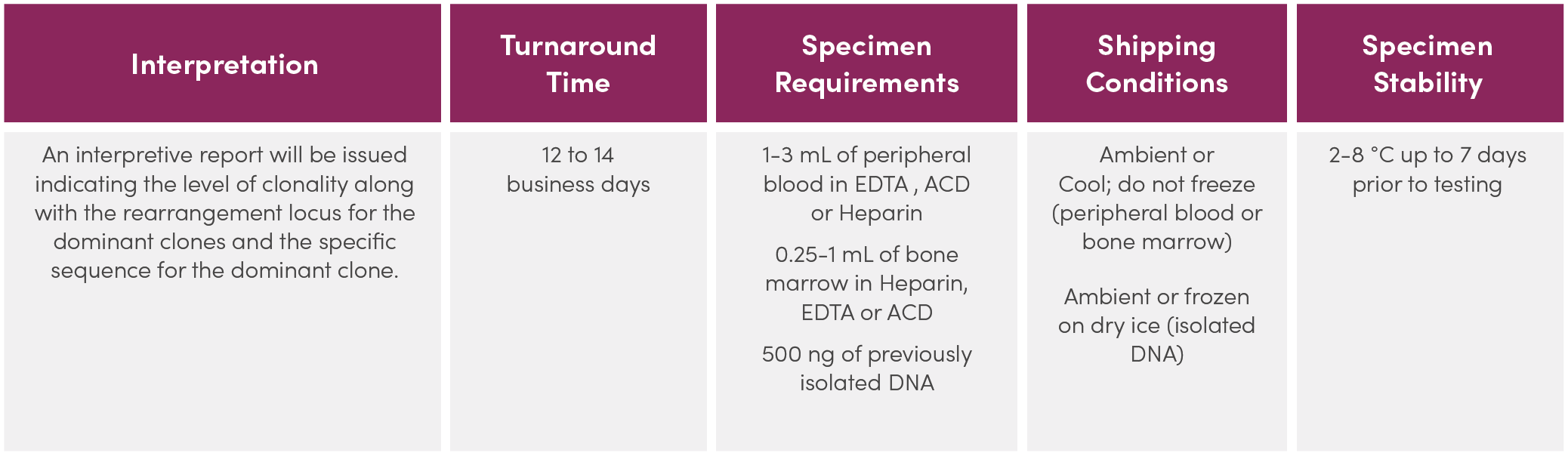
Service Details
-
Indications for Testing:
• Identify clonality in atypical lymphoproliferative disorders
• Support a differential diagnosis between reactive lesions and hematologic malignancies
• Assign presumptive lineage in mature monoclonal lymphoproliferative disorders
• Monitor and evaluate disease recurrence -
Service Description:
Legal Notice
Please contact Invivoscribe, Inc. for more information.
References
1. Tonegawa, S. (1983) Nature. 302:575-581.
2. Miller, JE et al. (2013, 2nd ed.) Springer Science & Business Media. 302.2.7.13 and 30.2.7.18.