 ITEM DISCONTINUED
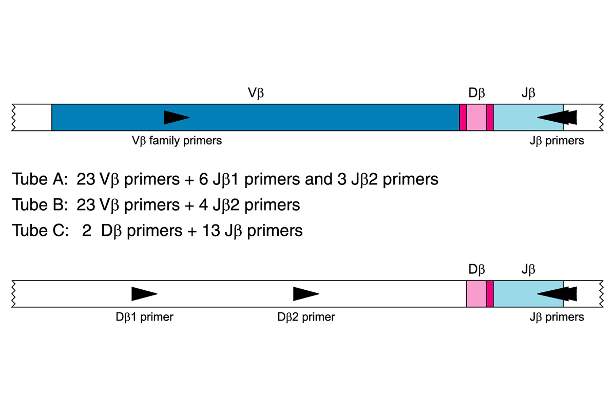
CAT #: 92050021
IdentiClone® TCRB Gene Clonality Assay MegaKit - ABI Fluorescence Detection
Intended Use
The IdentiClone® TCRB Gene Clonality Assay is an in vitro diagnostic product intended for PCR-based detection of clonal T-cell receptor beta chain gene rearrangements in patients with suspect lymphoproliferations and can be used to:
- Identify clonality in suspect lymphoproliferations
- Support a differential diagnsosis between reactive lesions and T-cell and some immature B-cell malignancies
- Determine lineage involvement in mature lymphoproliferative disorders
- Monitor and evaluate disease recurrence
Product Details
-
Summary and Explanation of the Test
Rearrangements of the antigen receptor genes occur during ontogeny in B and T lymphocytes. These gene rearrangements generate products that are unique in length and sequence for each cell. Therefore, polymerase chain reaction (PCR) assays can be used to identify lymphocyte populations derived from a single cell by detecting the unique V-J gene rearrangements present within these antigen receptor loci.1 This IdentiClone PCR assay employs multiple consensus DNA primers that target conserved genetic regions within the T-cell receptor beta chain gene. This test is used to detect the vast majority of clonal T-cell malignancies from DNA. Test products can be analyzed using a variety of detection formats, including gel and capillary electrophoresis.
Invivoscribe’s IdentiClone assays represent a simple approach to PCR-based clonality testing. These standardized assays were carefully optimized testing positive and negative control samples using multiplex master mixes. Assay development was followed by extensive validation including the testing of more than 400 clinical samples using Revised European/American Lymphoma (REAL) Classification. Testing was performed at more than thirty prominent independent testing centers throughout Europe in a collaborative study known as the BIOMED-2 Concerted Action.2
The ABI detection based assays cannot reliably detect clonal populations comprising less than 1% of the total lymphocyte cell population. Always interpret the results of molecular clonality tests in the context of clinical, histological and immunophenotypic data.
This test kit includes four (4) master mixes. TCRB Tubes A and B target framework regions within the variable region and the joining region of the TCR beta chain locus. TCRB Tube C targets the diversity and joining regions. Lastly, the Specimen Control Size Ladder master mix targets multiple genes and generates a series of amplicons of approximately 100, 200, 300, 400, and 600 base pairs to ensure that the quality and quantity of input DNA is adequate to yield a valid result. A single thermal cycler program and similar detection methodologies are used with all of our Gene Clonality Assays. This improves consistency and facilitates cross training on a broad range of different assays.
-
Principles of the Procedure
Polymerase Chain Reaction (PCR)
PCR assays are routinely used for the identification of clonal T-cell populations. These tests amplify the DNA between primers that target the conserved variable (V) regions and the conserved joining (J) regions (TCRB Tubes A and B), as well as the diversity (D) and joining regions (TCRB Tube C). These conserved regions lie on either side of an area within the V-J region where programmed genetic rearrangements occur during maturation of all B and T lymphocytes. The antigen receptor genes that undergo rearrangement are the immunoglobulin heavy chain and light chains in B-cells, and the T cell receptor genes in T-cells. Each B- and T-cell has a single productive V-J rearrangement that is unique in both length and sequence. Therefore, when DNA from a normal or polyclonal population is amplified using DNA primers that flank the V-J region, a bell-shaped curve (Gaussian distribution) of amplicon products within an expected size range is generated. On a gel, this distribution of products is observed as a smear. This Gaussian distribution reflects the heterogeneous population of V-J rearrangements. (In certain cases, where lymphocyte DNA is not present, no product is observed.) DNA from samples containing a clonal population yield one or two prominent amplified products (amplicons) within a diminished polyclonal background.
Since the antigen receptor genes are polymorphic (consisting of a heterogeneous population of related DNA sequences), it is difficult to employ a single set of DNA primer sequences to target all of the conserved flanking regions around the V-J rearrangement. N-region diversity and somatic mutation further scramble the DNA sequences in these regions. Therefore, multiplex master mixes, which target several FR regions, are required to identify the majority of clonal rearrangements. As indicated, clonal rearrangements are identified as prominent, single-sized products within the background of different-sized amplicon products that form a Gaussian distribution around a statistically favored, average-sized rearrangement.
Differential Fluorescence Detection
Differential fluorescence detection is commonly used to resolve the different-sized amplicon products using a capillary electrophoresis instrument. Primers can be conjugated with several different fluorescent dyes (fluorophors) so that they can produce different emission spectra upon excitation by a laser in the capillary electrophoresis instrument. In this manner, different fluorescent dyes can correspond to different targeted regions. This detection system results in unsurpassed sensitivity, single nucleotide resolution, differential product detection, and relative quantification. In addition, the use of agarose and polyacrylamide gels, as well as the use of carcinogens such as ethidium bromide, can virtually be eliminated. Further, differential detection allows accurate, reproducible and objective interpretation of primer-specific products and automatic archiving of data. Inter-assay and intra-assay reproducibility in size determination using capillary electrophoresis is approximately 1 to 2 nucleotides. This reproducibility and sensitivity coupled with the automatic archiving of specimen data allows for the monitoring, tracking, and comparison of data from individual patients over time.
-
Specimen Requirements
This assay tests genomic DNA (gDNA) from the following sources:
- 5 cc of peripheral blood, bone marrow biopsy, or bone marrow aspirate anti-coagulated with heparin or EDTA (stored at 2°C to 8°C and shipped at ambient temperature)
- Minimum 5 mm cube of tissue (stored and shipped frozen; or stored and shipped in RPMI 1640 at ambient temperature or on ice)
- 2 µg of DNA (stored at 2°C to 8°C and shipped at ambient temperature)
- Formalin-fixed paraffin embedded tissue or slides (stored and shipped at ambient temperature)
Disclaimer
This assay is based on the EuroClonality/BIOMED-2 Concerted Action BMH4-CT98-3936.
Legal Notice
For Legal Notices related to this product, visit: https://invivoscribe.com/legal-notices/