
CAT #: TRB-Clonality-Assay
TRB Clonality Assay
CONTACT US ⟶-
Description of Test:
For detection of the vast majority of TRB gene rearrangements, a multiplex master mix, targeting the Vß, Jß and Dß regions, is used for PCR amplification. Next-generation sequencing of the PCR products is used to identify DNA sequences specific to clonal gene rearrangements. Bioinformatics tools facilitate the characterization of sequences present at greater than 2.5% of the population. These sequences can be used to track specific clonal populations.
-
Overview:
The human T-cell receptor beta (TRB) gene locus on chromosome 7 (7q34) includes 64-67 variable (Vß) gene segments (belonging to 30 subgroups), 2 diversity (Dß) gene segments, and 13 joining (Jß) gene segments, spread over 685 kilobases, making this locus far more complex than others. Nevertheless, accurate molecular analysis of the TRB genes is an important tool for the assessment of clonality in suspected T-cell and some B-cell proliferations, as TRB gene rearrangements occur not only in almost all mature T-cell malignancies, but also in about one-third of precursor B-acute lymphoblastic leukemias (B-ALL).1
Lymphoid cells are different from the other somatic cells in the body, as during development the antigen receptor genes in lymphoid cells (including gene segments within the TRB locus), undergo somatic gene rearrangement.2
These developmentally regulated, programmed gene rearrangements generate combinations that are unique for each cell.1 Since leukemias and lymphomas originate from the malignant transformation of individual lymphoid cells, all leukemias and lymphomas generally share one or more cell-specific or “clonal” antigen receptor gene rearrangements. Clonality does not always imply malignancy; all results must be interpreted in the context of all of the other available diagnostic criteria. Tests that detect TRB clonal rearrangements can be used to help identify T-cell and certain B-cell malignancies.
CLINICAL UTILITY
T-cell receptor (TCR) gene rearrangement studies are often used in practice in conjunction with morphologic and immunophenotypic evaluation to distinguish T-cell lymphoma from reactive T-cell proliferations. They are particularly useful under the following conditions:
• Flow cytometric analysis on blood, bone marrow, nodal tissue/extra nodal tissue or body fluid (such as CSF, plural effusion, etc.) shows immunophenotypic abnormalities in the T-cells, but it is uncertain if the T-cells are truly clonal.
• Biopsy tissue shows atypical lymphoid infiltrate, and a clonal T-cell proliferation/T-cell neoplasm is suspected, such as biopsies of skin, lung, gastrointestinal tract, and nasal cavities. For skin biopsies, TCR gene rearrangement studies are particularly useful when distinguishing early evolving lymphoma (including early-stage mycosis fungoides) from some reactive conditions such as chronic dermatitis, drug reaction, autoimmune disease, viral infection, etc.
• Evaluating clonal relationships among different lesions, including multiple lesions from different locations and/or different time points.
While these genetic studies are essential for the differential diagnosis of T-cell lymphoma versus reactive T-cell proliferation sometimes, it is important to understand that the diagnosis cannot be based on molecular studies alone, but requires the careful correlation of clinical, morphologic and genetic features.
Service Details
-
Indications for Testing:
- Identify clonality in atypical lymphoproliferative disorders
- Support a differential diagnosis between reactive lesions and hematologic malignancies
- Assign presumptive lineage in mature monoclonal lymphoproliferative disorders
- Monitor and evaluate disease recurrence
-
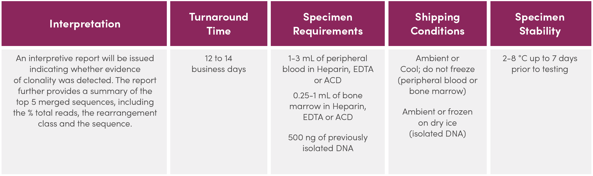
Service Description:
Legal Notice
Please contact Invivoscribe, Inc. for more information.
References
- Miller, JE et al. (2013, 2nd ed.) Springer Science & Business Media.2.7.13 and 30.2.7.18.
- Tonegawa, S. (1983) Nature. 302:575-581.